Smartphone Nosema Spore Analysis
Understanding Nosema Disease and Its Impact on Bees
Nosema disease, caused by the microsporidian parasites Nosema apis and Nosema ceranae , is a leading contributor to honeybee colony decline worldwide. These spores infect the digestive tracts of bees, impairing nutrient absorption and weakening immune systems. Infected colonies often exhibit reduced lifespan, lower honey production, and increased susceptibility to pesticides and other pathogens. The economic repercussions are significant: pollinators like honeybees are responsible for one-third of global food production, making Nosema a critical concern for agriculture and biodiversity.
Traditional Methods of Nosema Spore Analysis
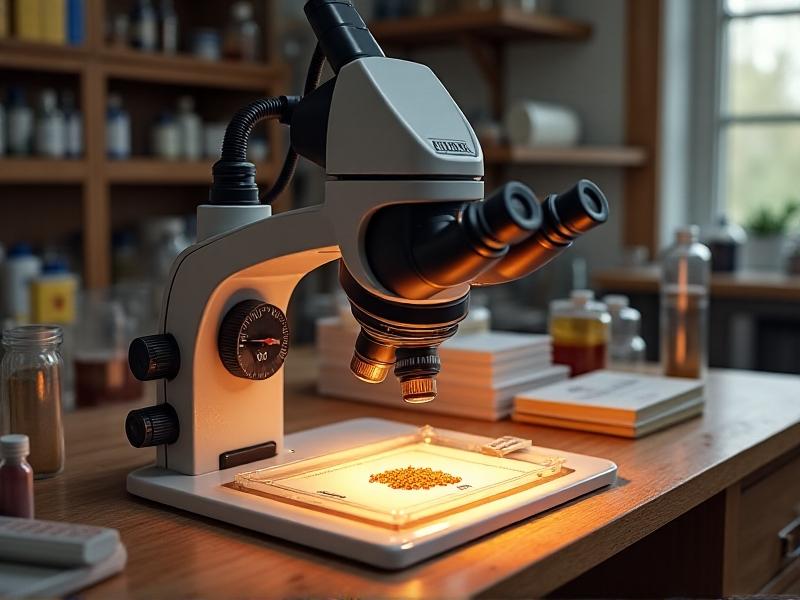
Historically, diagnosing Nosema has relied on laboratory-based microscopy. Technicians stain spore samples and manually count them under high-powered microscopes, a process requiring specialized training and equipment. While accurate, this method is time-consuming and inaccessible to field researchers or beekeepers in remote areas. Delays in diagnosis often lead to unchecked outbreaks, exacerbating colony losses. The need for rapid, on-site detection has driven innovation toward portable solutions, setting the stage for smartphone-based alternatives.
Emergence of Smartphone-Based Diagnostic Tools
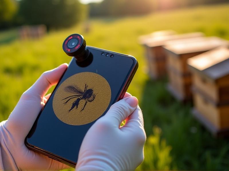
Smartphone microscopy leverages clip-on lenses or 3D-printed attachments to transform mobile devices into portable microscopes. These tools magnify spore samples up to 400x, capturing high-resolution images for analysis. Apps like BeeScan or SporeCam enable users to upload images, where built-in algorithms quantify spore concentrations. This approach democratizes diagnostics, allowing beekeepers to monitor hive health in real-time without laboratory access. The affordability—often under $50 per attachment—makes it a game-changer for resource-limited communities.
Technical Components of Smartphone Microscopy
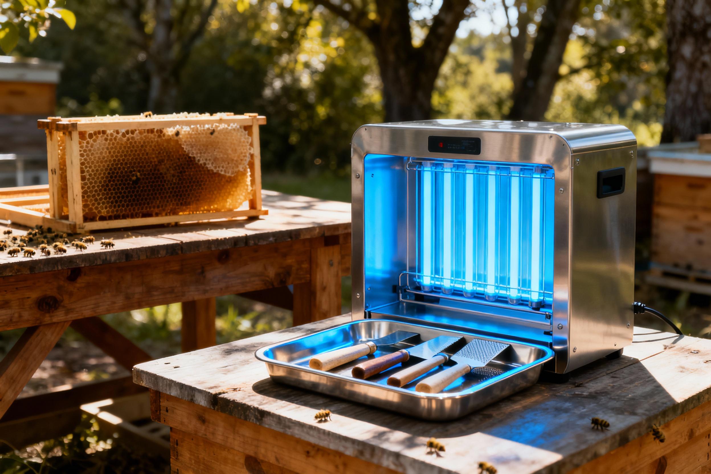
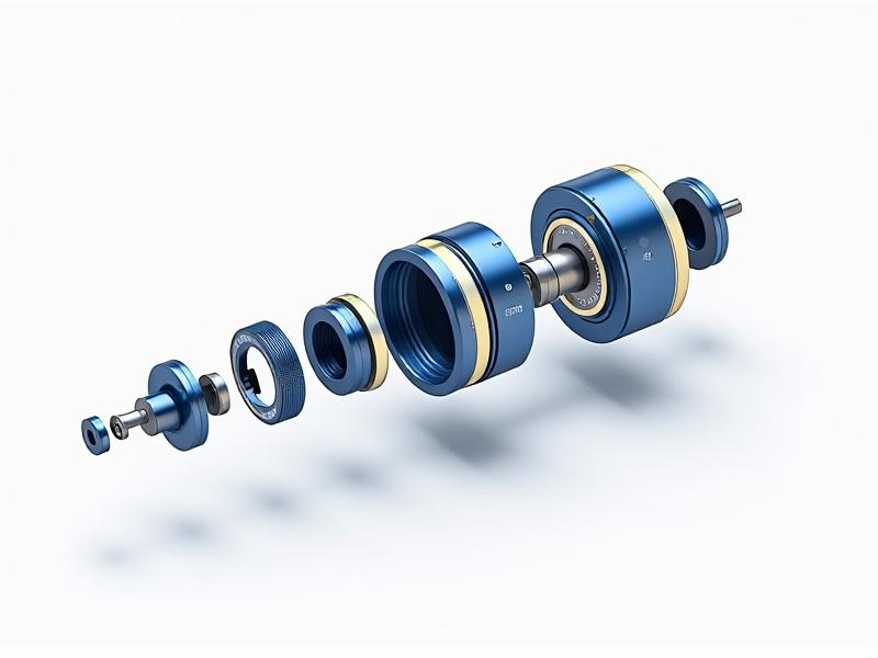
These systems combine hardware and software for end-to-end analysis. The hardware includes achromatic lenses for reduced chromatic aberration and adjustable LED lighting. Software components handle image stabilization, auto-focus, and background subtraction to enhance spore visibility. Open-source platforms like ImageJ have been adapted to process spore counts, while machine learning models trained on thousands of images improve detection accuracy. Cloud integration allows users to share data with researchers, creating crowdsourced maps of Nosema outbreaks.
AI and Machine Learning in Spore Detection
Convolutional neural networks (CNNs) now automate spore identification with over 95% accuracy. These models are trained on datasets comprising diverse spore morphologies, lighting conditions, and sample impurities. For instance, projects like HiveMind AI use transfer learning to adapt pre-trained models for specific regional spore variants. Real-time inference on edge devices eliminates cloud dependency, ensuring functionality in areas with poor connectivity. The result is a seamless workflow: capture, analyze, and act—all within minutes.
Case Studies: Successful Deployments in Apiaries
In 2022, a pilot program in Costa Rica equipped 150 beekeepers with smartphone kits. Over six months, participants reported a 40% reduction in colony losses compared to control groups. Similarly, Vermont’s Beekeepers Association integrated mobile diagnostics into their annual health audits, slashing lab turnaround times from weeks to hours. These successes highlight how accessible technology can bridge the gap between research and grassroots conservation efforts.
Advantages Over Conventional Laboratory Techniques
Beyond speed and cost, mobile analysis preserves sample integrity by minimizing transport delays. Traditional methods risk spore degradation during shipping, especially in高温 climates. Smartphone workflows also generate digital records, enabling longitudinal tracking of hive health. For researchers, this data trove offers unprecedented insights into spore proliferation patterns and the efficacy of miticides or dietary supplements.
Challenges in Accuracy and Standardization
Variability in smartphone cameras and lighting conditions can skew results. A 2023 study found that lower-end devices struggled with spore differentiation in debris-heavy samples. Efforts to standardize protocols—such as uniform staining methods and calibration slides—are underway. Collaborative initiatives like the Global Nosema Consortium aim to establish certified training programs and device benchmarks to ensure reliability across platforms.
Future Directions: Enhancing Accessibility and Precision
Next-gen attachments may incorporate Raman spectroscopy for molecular-level spore analysis. Meanwhile, satellite-connected devices could relay hive data to centralized dashboards, alerting beekeepers to regional outbreaks. Partnerships with telecom companies aim to subsidize kits in developing regions, fostering global adoption. As AI models evolve to predict colony collapse from early spore counts, proactive interventions could redefine sustainable apiculture.
Empowering Beekeepers Through Mobile Technology
The shift from reactive to preventive beekeeping hinges on accessible diagnostics. By equipping individuals with smartphone tools, communities gain agency in combating Nosema. Workshops and citizen science projects further demystify the technology, fostering a new generation of tech-savvy stewards. As these innovations scale, they promise not only healthier bees but also more resilient ecosystems—one hive at a time.